When you sit in the chair for a checkup, the dentist can see a lot. Chips, stains, swollen gums, the edges of old fillings. But most of each tooth sits below the gumline or hidden between its neighbours, and some of the most common problems start exactly there. Dental X-rays let us see those parts. At Sherwood Dental in Kitchener, they’re one of the most useful tools we have for catching problems early and planning treatment properly, and patients often ask what we’re actually looking at. This post explains it.
What an X-ray can show that an exam can’t
X-rays pass through soft tissue easily and are partly blocked by denser material like enamel, bone and metal. The result is an image where healthy tooth structure looks light and anything less dense, like decay or infection, shows up darker. That contrast reveals things we’d otherwise miss:
- Cavities between teeth. The surfaces where two teeth touch are invisible during a normal exam. Decay often starts here and can grow quietly for a long time.
- Decay under existing fillings or crowns. A filling can look fine on top while the tooth underneath is breaking down.
- Bone loss from gum disease. Gum disease slowly destroys the bone around teeth. X-rays show how much bone is left and whether it’s changing.
- Infections at the root tip. An abscess can form at the end of a root, sometimes without pain at first.
- Impacted or extra teeth. Wisdom teeth that are stuck under the gum, or teeth that never came in, are easy to spot.
- Cysts and other unusual findings. These are rare, but X-rays are often how they’re first noticed.
The main types of dental X-rays
“X-ray” covers a few different images, each taken for a different reason. You might have one or several depending on what we need to see.
Bitewings
These are the small images taken while you bite down on a little tab or holder. Each one shows the upper and lower back teeth on one side. Bitewings are the go-to for finding cavities between teeth and checking bone levels.
Periapicals
A periapical image shows one or two teeth from the crown all the way to the tip of the root. We use these when a specific tooth is bothering you, when we suspect an infection, or before and during a root canal.
Panoramic X-rays
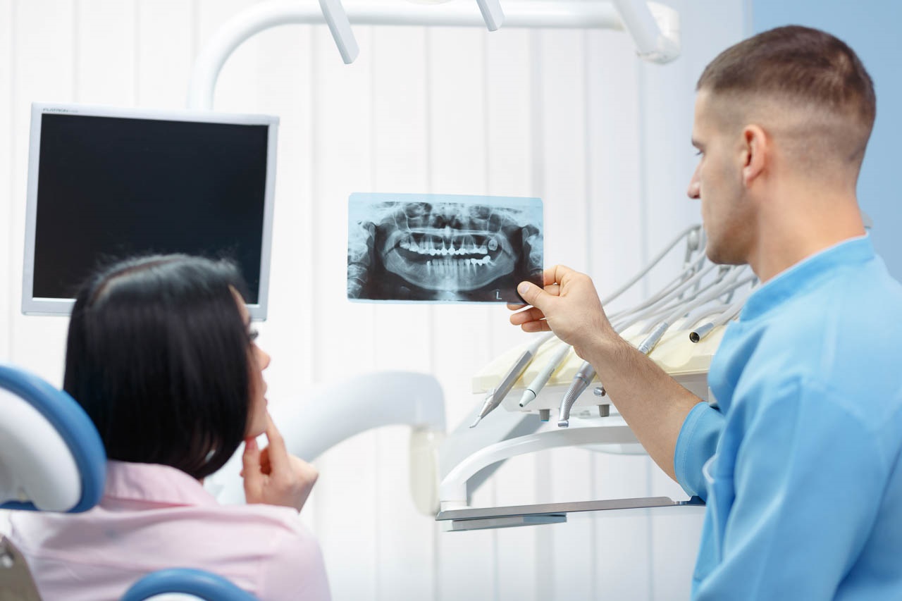
A panoramic image wraps around your whole jaw in a single picture. The machine moves around your head while you stand or sit still. It’s useful for seeing wisdom teeth, the jaw joints and the overall layout of your teeth and jaws, though it doesn’t show fine detail as well as bitewings.
How X-rays shape treatment at Sherwood Dental
Finding a problem is only half the job. X-rays also help us decide what to do about it, and how.
Take a cavity. On an X-ray we can see how deep it goes. A shallow one needs a simple filling. One that’s close to the nerve might need a different approach, and one that has reached the nerve may need a root canal. Knowing this ahead of time means fewer surprises halfway through an appointment.
For gum disease, comparing images over the years shows whether bone levels are stable or dropping, which tells us whether home care and regular cleanings are working or whether more treatment is needed.
Before an extraction, X-rays show the shape and position of the roots and how close they sit to nerves or the sinus. For anyone considering a replacement tooth down the road, they help us see whether there’s enough bone to support it.
Looking at your X-rays together
X-rays can look like a blur of grey shapes if no one walks you through them. We’re happy to put your images up and point out what we’re seeing: a dark shadow between two molars, the outline of an old filling, the line of bone along your roots. Seeing it yourself makes it much easier to understand why we’re suggesting a filling now, or why we just want to keep an eye on a spot for another six months. If you’d like to look, just ask at your appointment.
How often do you need dental X-rays?
There’s no single schedule that fits everyone. We take X-rays when the information is likely to change what we do, and we base the timing on your own history. In general:
- New patients often need a set so we have a starting point, unless recent images can be sent from a previous office
- Adults with healthy teeth and few fillings may only need bitewings every couple of years
- People who get cavities easily, or who are being treated for gum disease, may need them more often
- Children’s X-rays are timed around their growth and the arrival of adult teeth
- Any time a tooth hurts or something looks wrong, we may take an image of that area
If you’re not sure why an X-ray is being recommended, ask. We’re always happy to explain what we’re looking for.
What about radiation?
This is a fair question and one we hear often. Dental X-rays use a very small amount of radiation, much less than many medical imaging tests. You’re also exposed to natural background radiation every day just from living on Earth, and a routine set of dental X-rays adds only a small amount on top of that.
Even so, we follow the principle of keeping exposure as low as reasonably possible. That means not taking images out of habit, only taking the ones that will help, and using protective measures where they’re appropriate. If you’re pregnant or think you might be, please tell us before any X-rays are taken. In most cases routine images can wait, and urgent ones can be taken with extra care.
Tips for an easier X-ray appointment
Most people find X-rays quick and easy, but a few things help:
- If you have a strong gag reflex, let us know. Breathing slowly through your nose and keeping your mind on something else really does help, and we can adjust how we position the sensor.
- Remove earrings, glasses and anything metal around your head and neck if we ask, since metal can block part of the image.
- Hold still for the few seconds while the image is taken. Moving can blur it and mean a retake.
- If you’ve had X-rays at another office recently, tell us. We can often request them instead of taking new ones.
Questions about dental X-rays
Can I refuse X-rays?
You can, and we’ll respect that. We’ll explain what we might miss without them so you can make an informed choice. Some treatment, such as a root canal, can’t safely be done without an image of the tooth.
Do X-rays hurt?
No. The worst part for most people is the sensor or film feeling a bit awkward in the mouth for a few seconds.
Are X-rays covered by insurance?
Most dental plans cover diagnostic X-rays, though they often limit how frequently. Our front desk can help you check your plan.
Book your checkup with Sherwood Dental in Kitchener
If you’re due for a checkup, or a tooth has been bothering you and you want to know what’s going on beneath the surface, call Sherwood Dental at 519-576-2170. We’re at 501 Krug St, Unit 113, in Kitchener, and we’ll be glad to explain anything we see on your X-rays.
The main types of dental X-rays
Bitewings, periapicals and panoramic images
Bitewing X-rays show the upper and lower back teeth together and are the best way to find cavities between teeth. A periapical X-ray shows one or two teeth from crown to root tip, which helps when a tooth is sore or a root canal is being considered. A panoramic X-ray captures both jaws in one image, so it is useful for wisdom teeth and for checking the jaw bone.
How often you need them
There is no fixed schedule for everyone. Patients with healthy teeth and few fillings may only need bitewings every year or two, while people who get cavities often or have gum problems may need them more often. Your dentist bases the timing on your own history.